PRP Preparation Methods: Why the Protocol Determines the Product.
Centrifugation speed, leukocytes, and platelet quality explained
Your doctor prescribed PRP. Or maybe you heard about it in a waiting room. Either way, almost nobody explained the one thing that makes all the difference: not just what PRP is, but how it is prepared.
Platelet-rich plasma is a concentrate made from your own blood. A small volume of venous blood is collected, placed in a centrifuge, and the result is a biologically active product rich in platelets and growth factors. That sounds straightforward. It is not.
A 2017 systematic review published in the Journal of Bone and Joint Surgery analyzed the clinical orthopedic literature on PRP and found that fewer than 10% of studies clearly described the preparation protocol used. Most published data on PRP cannot be compared between studies, because every lab prepares something different under the same name.
This article explains what physically happens inside the tube during centrifugation, which variables determine the final product composition, and why choosing the wrong protocol can change the therapeutic result entirely.
What Happens Inside the Tube During Centrifugation
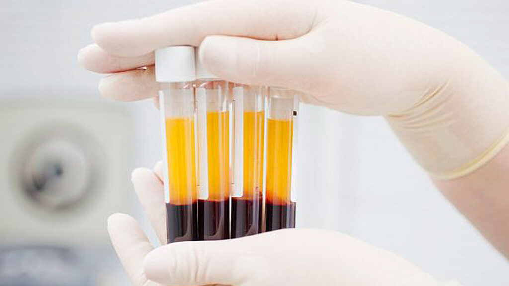
When whole blood enters a centrifuge, its components separate by density. Heavier particles migrate toward the bottom of the tube; lighter ones stay near the top. This process is called differential centrifugation, and it is the same principle used in laboratories worldwide to separate blood fractions.
The three main blood components have different densities. Red blood cells (RBCs) are the densest, at 1095 to 1100 kg/m³, and settle to the bottom. White blood cells (WBCs) have a density of 1055 to 1085 kg/m³, and form a thin layer called the buffy coat — the whitish layer between the yellow plasma and the red cells. Platelets are the least dense of all, at 1040 to 1065 kg/m³, and remain suspended in the plasma above.
In practice, the density difference between leukocytes and red blood cells is small — but it is that small difference that makes precise separation possible or impossible. There is another complicating factor: red blood cells outnumber white blood cells by more than a thousand to one. Five million RBCs versus 5,000 WBCs per microliter of blood. That ratio affects how well any centrifuge separates these populations.
A 2020 study published in BMC Oral Health by Miron, Zhang, and colleagues analyzed 24 different centrifugation protocols, measuring cell distribution layer by layer — each milliliter sampled separately — across 960 complete blood count analyses. Researchers used 6 centrifugal forces (100, 200, 400, 700, 1000, and 1200g) combined with 4 centrifugation times (3, 5, 8, and 12 minutes) on a horizontal centrifuge.
The clearest finding: platelets distributed into the upper plasma layers easily because of their lower density. Leukocytes tended to accumulate in the buffy coat. Getting both cell types evenly distributed in the upper plasma — which is what clinicians actually want — required precise protocol calibration. Notably, centrifugation time had a greater impact on final cell layer separation than speed did. That is counterintuitive, and it explains many of the inconsistencies in published clinical results.
KEY DATA: Blood cell density in whole blood • Platelets: 1040-1065 kg/m3 (least dense — stay in upper plasma) • White blood cells: 1055-1085 kg/m3 (intermediate — tend toward buffy coat) • Red blood cells: 1095-1100 kg/m3 (most dense — settle to the bottom) • Numerical ratio: 5 million RBCs vs 5,000 WBCs per microliter of blood |
Four Types of Platelet Concentrates
The term PRP covers very different products. In 2009, researcher Dohan Ehrenfest proposed a systematic classification of platelet concentrates based on two variables: leukocyte content and fibrin architecture. That classification remains the standard reference in the literature today.
The classification identifies four categories:
- P-PRP (Pure Platelet-Rich Plasma): high platelet concentration, no leukocytes, low-density fibrin network. Typically produced with double centrifugation at lower speeds.
- L-PRP (Leukocyte and Platelet-Rich Plasma): platelets and leukocytes, with a low-density fibrin network. This category has the largest number of commercial systems available.
- P-PRF (Pure Platelet-Rich Fibrin): platelets without leukocytes, high-density fibrin network. Exists only as a solid gel — it cannot be injected.
- L-PRF (Leukocyte and Platelet-Rich Fibrin): platelets and leukocytes, high-density fibrin. The second-generation form prepared without anticoagulants, which forms a solid membrane after centrifugation.
This classification is not just academic. Different tissues respond differently to leukocyte content. A 2021 narrative review in EFORT Open Reviews reported that leukocyte-rich PRP appears more effective in tendinopathies, while leukocyte-poor PRP produces better outcomes in cartilage pathology. Leukocytes release proteases and pro-inflammatory cytokines that stimulate regeneration in tendons, but in cartilage the same inflammatory environment can accelerate tissue damage.
A 2016 study in the Journal of Orthopaedic Science (Kobayashi et al.) compared three preparations and found dramatically different leukocyte concentrations: 14.9 × 10³/μl in leukocyte-rich PRP versus 0.2 × 10³/μl in pure PRP. In both cases, platelet concentration correlated positively with all growth factors measured.
Two patients can receive an injection called “PRP” and receive products with biologically opposite compositions. For more on how PRP is used in specific conditions like knee osteoarthritis, see our article on platelet-rich plasma for knee pain.
Single Centrifugation Versus Double Centrifugation
PRP is prepared primarily with two methods: double centrifugation and the buffy coat method. The difference is not only technical, it changes the composition of the final product.
In the double centrifugation method, whole blood is centrifuged once at low speed (soft spin) to separate red blood cells from plasma. After this first spin, the upper plasma layer — containing platelets and leukocytes — is transferred to a second tube and centrifuged again at higher speed (hard spin) to concentrate platelets at the bottom. The platelet pellet is then resuspended in a small volume of plasma. One known limitation: approximately 20% of platelets remain adsorbed to the residual red blood cells in the transferred plasma and are not recovered.
In the buffy coat method, whole blood is centrifuged at high speed in a single step. The buffy coat layer — rich in platelets and leukocytes — is manually collected or filtered. The advantage is higher leukocyte concentration in the final product. The disadvantage is the technical difficulty of separating this thin layer without contaminating it with the underlying red blood cells.
Double centrifugation produces the highest yield. A study by Amanda et al., cited in the Dhurat and Sukesh review (Journal of Cutaneous and Aesthetic Surgery, 2014), demonstrated that processing 3.5 mL of blood at 100g for 10 minutes (first spin) then 400g for 10 minutes (second spin) achieved platelet recovery of 70% to 80%, with a concentration five times the baseline value in whole blood.
One underappreciated factor concerns the time between blood collection and centrifugation. For PRF — prepared without anticoagulants — blood begins to clot immediately after collection. The 2020 Miron et al. study emphasizes that the clinician must begin centrifugation immediately after collection, because fibrin formation alters cell distribution inside the tube.
For more on how exercise before blood draw can improve platelet count and PRP quality, see our article on how exercise boosts platelet count and PRP quality.
The Three Variables That Change Everything: Speed, Time, and Temperature
Three physical variables determine the final quality of PRP: relative centrifugal force (RCF), centrifugation time, and temperature. Each variable acts independently and interacts with the others in non-linear ways.
Relative centrifugal force (RCF). RCF is not the same as rotations per minute (RPM). The same machine with rotors of different radius produces different g-forces at the same RPM. The formula is: g = (1.118 × 10⁻⁵) × R × S², where R is the rotor radius in centimeters and S is the speed in RPM. Many published studies that report only RPM without specifying rotor radius are not reproducible by other labs.
The Miron et al. study identified optimal ranges for two distinct products:
- Liquid PRP (injectable i-PRF): the optimal protocol is 200g for 5 minutes. This produces the highest concentration of platelets and leukocytes in a reduced plasma volume.
- Solid PRF: the optimal range is 400g to 700g for 8 minutes. At 700g for 8 minutes, platelet yield reached 96% of the total, with even distribution across the upper plasma layers.
Protocols below 200g did not effectively separate cells. Protocols above 1000g for 5 minutes, or 12-minute protocols at 400g or higher, pushed leukocytes exclusively into the buffy coat, with minimal presence in the upper plasma.
Centrifugation time. Longer centrifugation improves total yield (more cells recovered) but reduces concentration, because the final plasma volume is larger. Beyond 8 minutes, leukocytes systematically migrate toward the buffy coat. This can be used deliberately: if the goal is leukocyte-poor PRP — preferred for cartilage pathology — extending centrifugation time is a strategic choice.
Temperature. Platelets activate prematurely when exposed to inadequate temperatures, releasing growth factors before injection. The American Association of Blood Banks recommends 21-24°C. Many researchers use 12-16°C for better platelet viability. A 2013 study by Amable et al. in Stem Cell Research & Therapy optimized a protocol at 300g for 5 minutes at 12°C for the first spin, followed by 700g for 17 minutes for the second spin, achieving platelet concentrations between 5.4 and 7.3 times the baseline value.
PRACTICAL NOTE: Standard diagnostic centrifuges are not designed for PRP preparation. They lack precise temperature control and often cannot reach the required speeds reproducibly. A refrigerated tabletop centrifuge designed for blood components is the most reliable solution for a controlled protocol. |
Horizontal Versus Fixed-Angle Centrifuge
The shape of the centrifuge changes how cells physically separate inside the tube. Most labs use fixed-angle centrifuges: the tube is tilted at a fixed angle during rotation, and centrifugal forces push cells toward the distal wall of the tube — the wall facing outward from the machine. Cells accumulate along that wall, then slide toward the bottom.
This creates two problems. First, cell layer separation occurs at an angle rather than horizontally. The boundary between plasma and red blood cells is not perpendicular to the tube axis, and separation is less distinct. Second, cells colliding with the distal wall experience mechanical stress (shear stress) that can damage them or activate them prematurely.
A horizontal centrifuge with a swing-out bucket rotor solves both problems. The tube moves to a fully horizontal position during rotation, with the tube bottom pointing outward. Separation occurs perpendicular to the tube axis, cell layers form horizontally, and the distance between the minimum centrifugal force point (RCF-min, near the center of rotation) and the maximum force point (RCF-max, at the tube bottom) is maximized.
This gap between RCF-min and RCF-max is critical: it is what allows clean layer separation. In a fixed-angle centrifuge, this differential is smaller and the resulting separation is less defined.
The Miron et al. 2020 study used an Eppendorf 5702 horizontal centrifuge and reported up to 3.5-fold improvements in platelet and leukocyte concentration compared to fixed-angle centrifuges used in standard L-PRF protocols. A previous paper from the same group showed that fixed-angle centrifugation produced uneven cell concentrations, with most cells found on the distal side of PRF membranes.
Under older i-PRF protocols on fixed-angle centrifuges (approximately 60g for 3-4 minutes), the cell concentration increase was only 33% above whole blood — insufficient for many clinical applications. The Miron study confirmed that protocols at 100g or below were ineffective regardless of centrifuge type.
Individual patient variability is another underappreciated factor. The Miron study centrifuged samples with the same protocol (700g for 5 minutes) from three different donors and found plasma volume differences exceeding 50%. Donor 1 produced approximately 3 mL of plasma; donor 3 produced approximately 5 mL with the identical protocol. This variability comes from individual hematocrit levels — the ratio of red blood cell volume to total blood volume.
Women and older patients typically have lower hematocrit, which accelerates cell layer separation during centrifugation, producing more plasma but with lower cell concentration per milliliter. A protocol optimized for a healthy adult male may not be optimal for a 65-year-old woman. Developing hematocrit-adjusted protocols is one of the open research questions in this field.
For more on how PRP is applied in sports injuries and joint pain, read our article on platelet-rich plasma for joint pain and sports injuries.
What This Means for You
Two clinics offering “PRP” can deliver completely different products. The difference is not in the name of the treatment — it is in every procedural choice: the anticoagulant used for blood collection, centrifuge type, speed, time, temperature, number of spins, and how the final product is collected and administered.
This does not mean PRP does not work. It means a specific type of PRP works for specific conditions, and a different type works for others. Leukocyte-poor PRP produces better outcomes in cartilage osteoarthritis, while leukocyte-rich PRP performs better in tendinopathies. To interpret these results, clinicians and patients need to know exactly what was prepared.
Preparation variability is the main reason PRP clinical results appear contradictory in the literature. Not because the product fails, but because studies are frequently comparing different products under the same name.
If you are evaluating PRP treatment, ask your doctor which protocol they use: what type of centrifuge, how many g and for how many minutes, whether the product is leukocyte-rich or leukocyte-poor, and whether they use a commercial kit or a custom protocol. Those questions are not technical details. They are the difference between a treatment matched to your tissue and one based on what the lab happens to stock.
For a broader overview of PRP’s role in regenerative medicine, see our article on platelet-rich plasma and its hidden healing power.
References
- Miron RJ, Chai J, Fujioka-Kobayashi M, Sculean A, Zhang Y. Evaluation of 24 protocols for the production of platelet-rich fibrin. BMC Oral Health. 2020;20(1):310.
- Dhurat R, Sukesh MS. Principles and methods of preparation of platelet-rich plasma: a review and author’s perspective. J Cutan Aesthet Surg. 2014;7(4):189-97.
- Dohan Ehrenfest DM, Rasmusson L, Albrektsson T. Classification of platelet concentrates: from pure platelet-rich plasma (P-PRP) to leucocyte- and platelet-rich fibrin (L-PRF). Trends Biotechnol. 2009;27(3):158-67.
- Chahla J, Cinque ME, Piuzzi NS, et al. A call for standardization in platelet-rich plasma preparation protocols and composition reporting: a systematic review of the clinical orthopaedic literature. J Bone Joint Surg Am. 2017;99(20):1769-79.
- Mazzocca AD, McCarthy MB, Chowaniec DM, et al. Platelet-rich plasma differs according to preparation method and human variability. J Bone Joint Surg Am. 2012;94(4):308-16.
- Kobayashi Y, Saita Y, Nishio H, et al. Leukocyte concentration and composition in platelet-rich plasma influences the growth factor and protease concentrations. J Orthop Sci. 2016;21(5):683-9.
© 2026 Alice & Marcus Guimarães. Tutti i diritti riservati. Questo sito è stato creato con WordPress.